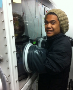
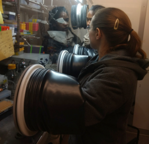
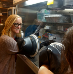
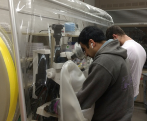
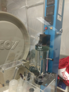
Oxygen sensitive proteins are purified in an MBraun anaerobic chamber located in our cold room. A Bio-Rad NGC FPLC is integrated into this glove box, allowing us to use any column we need for anaerobic purifications. There is also a microscope for setting up crystal trays and looping at 4°C.
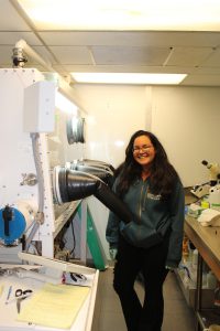
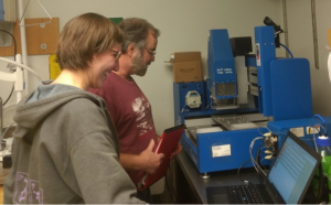
Crystallization conditions for oxygen sensitive proteins are screened using a Mosquito drop-setting robot in an MBraun Chamber housed in the Laboratory for Anaerobic Crystallography (LAC@MIT) on the 5th floor of building 68. The trays are imaged in a Formulatrix hotel also located in the chamber.
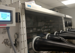
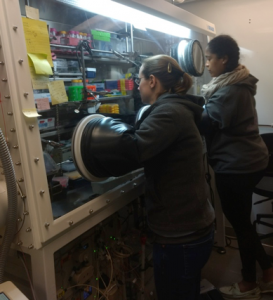
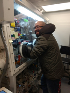
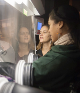
After initial conditions are identified we can optimize the crystals in a second MBraun in the LAC@MIT. This box has a microscope for examining trays and looping and can also be used for kinetic assays.
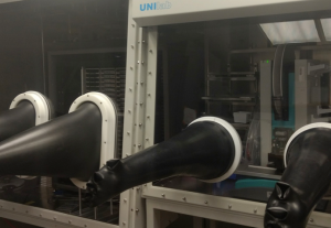
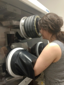
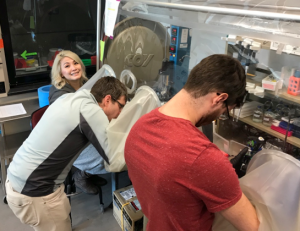
Most of our anaerobic looping is done in a Coy anaerobic tent in our labs on the 6th floor. Just before a synchrotron trip this tent can get a bit crazy!
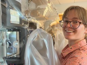
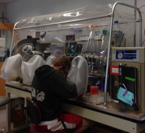
We have a second Coy anaerobic tent that houses a Vitrobot for prepping anaerobic cryo-EM grids. (Aerobic cryo-EM samples can be prepared on the EM facility Vitrobot plunger or with the chameleon for blot-free grid preparation). We bring our ITC inside this tent when we want to determine binding constants for anaerobic proteins. This Coy is also used for growing obligate anaerobic bacterial species.
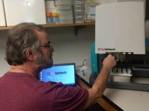
We screen crystallization conditions for non-oxygen sensitive proteins using either the Phoenix or NT8 robots in the Structural Biology Service Center located just below us. Trays can be imaged at either 18°C or 4°C in Formulatrix imaging hotels. Promising conditions can be further optimized on the Phoenix or NT8 using custom screens created on the Dragonfly robot in the Center.
Building 68 has a home source X-ray diffraction set-up so we can screen our crystals before sending them to the synchrotron. This set-up also allows us to collect full datasets at home, including single-wavelength anomalous datasets. We house our UV-Vis microspectrophotometer in the same room so that we can collect spectra on crystals before and after they are in the beam. We send our crystals to APS, SSRL.
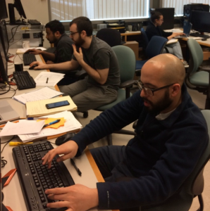
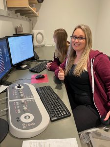
We have multiple Linux workstations equipped with 3D graphics cards and monitors so we can easily alternate refinement and inspection of our structures in 3D in COOT. We have computational resources in building 68 and at the Holyoke computing facility (MGHPCC) for processing cryo-EM datasets. We are members of the SBGrid consortium and they ensure that we have the latest version of all of the structural biology software we use installed and ready to use.
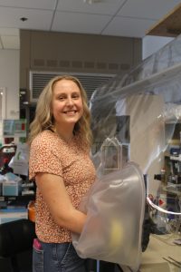
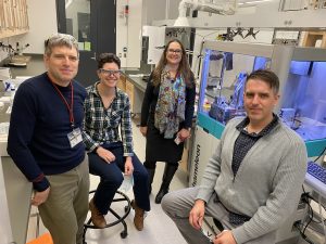
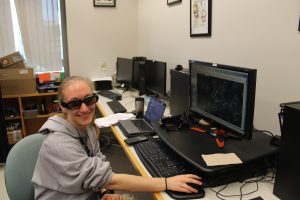
For cryo-EM data collection, we have access to a Talos Arctica microscope and a Titan Krios microscope, which are housed in the cryo-EM facility in the MIT.nano building. Our former postdoc Ed Brignole (pictured) set up the facility.
Other facilities we use on campus to characterize our proteins include:
Department of Chemistry Instrumentation Facility (NMR, EPR, LC-MS)
Biophysical Instrumentation Facility (Analytical Ultracentrifugation, circular dichroism spectroscopy, dynamic light scattering, mass photometer, NanoTemper Prometheus Panta nanoDSFl Octet R8 Bio-Layer Interferometry)
Center for Environmental Health Bioimaging and Chemical Analysis Core Facility (MS, LC-MS)
Swanson Biotechnology Center Biopolymers and Proteomics Core Facility (protein and peptide MS)
Francis Bitter Magnet Laboratory (NMR, EPR)